
Articles on Physiotherapy Treatments & Musculoskeletal Conditions
This Physiotherapy Articles portal is dedicated to providing clear information on the latest physiotherapy conditions and treatment for online users.
Getting better is not just your physiotherapists, instructors or trainers’ responsibility. It’s is also yours, your care-takers and loved one’s. It is a collaborative effort that require everyone’s active understanding and participation.
Here at Core Concepts, we’re about overcoming barriers and expanding human potential. We’re about enriching the lives of our clients and inspiring them to get back to their best.

Strength is a Shield, Not a Suit of Armor: 3 Reasons Why the Fittest Still Get Injured
Date
20 May 2026
Strength and fitness are significant protective factors, but they do not provide absolute immunity to...

Shortening rehabilitation time with psychosocial factors
Date
13 April 2026
Recovering from an injury involves more than just repairing muscles, joints, or tissues. A person’s...
Plantar Fasciitis Treatment: Persistent Heel Pain — and What Actually Works
Date
27 February 2026
Plantar Fasciitis Treatment for Persistent Heel Pain Plantar fasciitis is one of the most common...
That Pain Running Down Your Leg Could Be From Your Hip, Not Your Spine
Date
26 September 2025
Referred Leg Pain: Why Your Hip Might Be the Real Culprit Pain travelling down your...

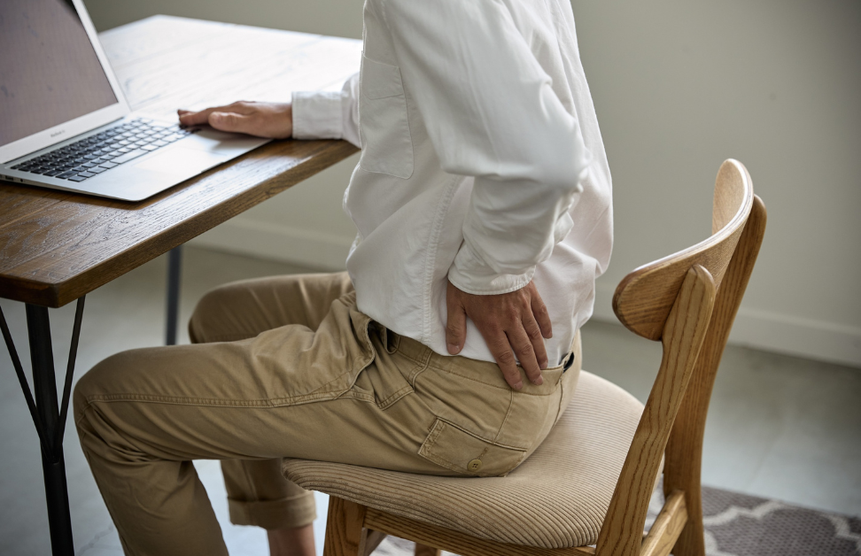
Could your desk ergonomics be hindering your climbing progress?
Date
19 June 2025
Climbing is a high-impact sport that requires strong muscles and healthy joints for longevity. Poor...

Solving The Puzzle of Persistent Pain: Why Some Recover While Others Suffer Chronically
Date
5 June 2025
The Chronic Pain Puzzle: Understanding Why Some People Heal Faster Beyond tissue damage, a powerful...

Rest Alone Isn’t Always the Answer for Plantar Fasciitis
Date
7 May 2025
Why Rest Alone Isn’t the Solution for Plantar Fasciitis Many people with heel pain instinctively...

Biceps Tendon and Shoulder Pain: A Gym-Goer’s Guide
Date
21 March 2025
That sharp shoulder pain at the gym might not be what you think - it...

Neglecting Ankle Sprains, could cause knee and hip issues in the future
Date
6 February 2025
The Kinetic Chain: How Ankle Sprains Impact Your Knees and Hips Ankle sprains are common...
