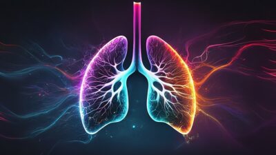
How Gut Bacteria Affect Lung Fibrosis
In Aging Cell, researchers have described how a strain of Lactobacillus gut bacteria sends chemical signals that enter the bloodstream and decrease fibrosis in the lungs. The gut-lung axis The


In Aging Cell, researchers have described how a strain of Lactobacillus gut bacteria sends chemical signals that enter the bloodstream and decrease fibrosis in the lungs. The gut-lung axis The
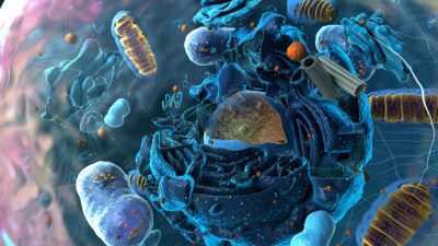
Researchers have explained how a protein found in both yeast and humans facilitates the destruction of the core protein responsible for Parkinson’s disease. An aggregate that impairs clearance The loss
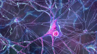
According to a new study, a special protein disposal system, currently found only in neurons, is linked to central hallmarks of Alzheimer’s disease [1]. The membranal proteasome Alzheimer’s disease is


The Longevity Investor Network (LIN) was created to help bridge the gap between promising longevity startups and the investors capable of helping them scale. Through curated monthly pitch sessions, educational

[Mountain View, September 17, 2025] — Lifespan Research Institute (LRI) today announced the launch of the Public Longevity Group (PLG), a new initiative focused on bridging the cultural gap between

Mountain View, California — Lifespan Research Institute, a nonprofit leader in longevity science and advocacy, announces the launch of the Lifespan Alliance, a sponsorship initiative uniting mission-driven companies and visionary
